CMOS MEA for high-throughput, multi-modal cell interfacing
This compact integrated micro-electrode array for intra- and extracellular electrical cell interfacing is a game-changer for disease modeling and drug development.
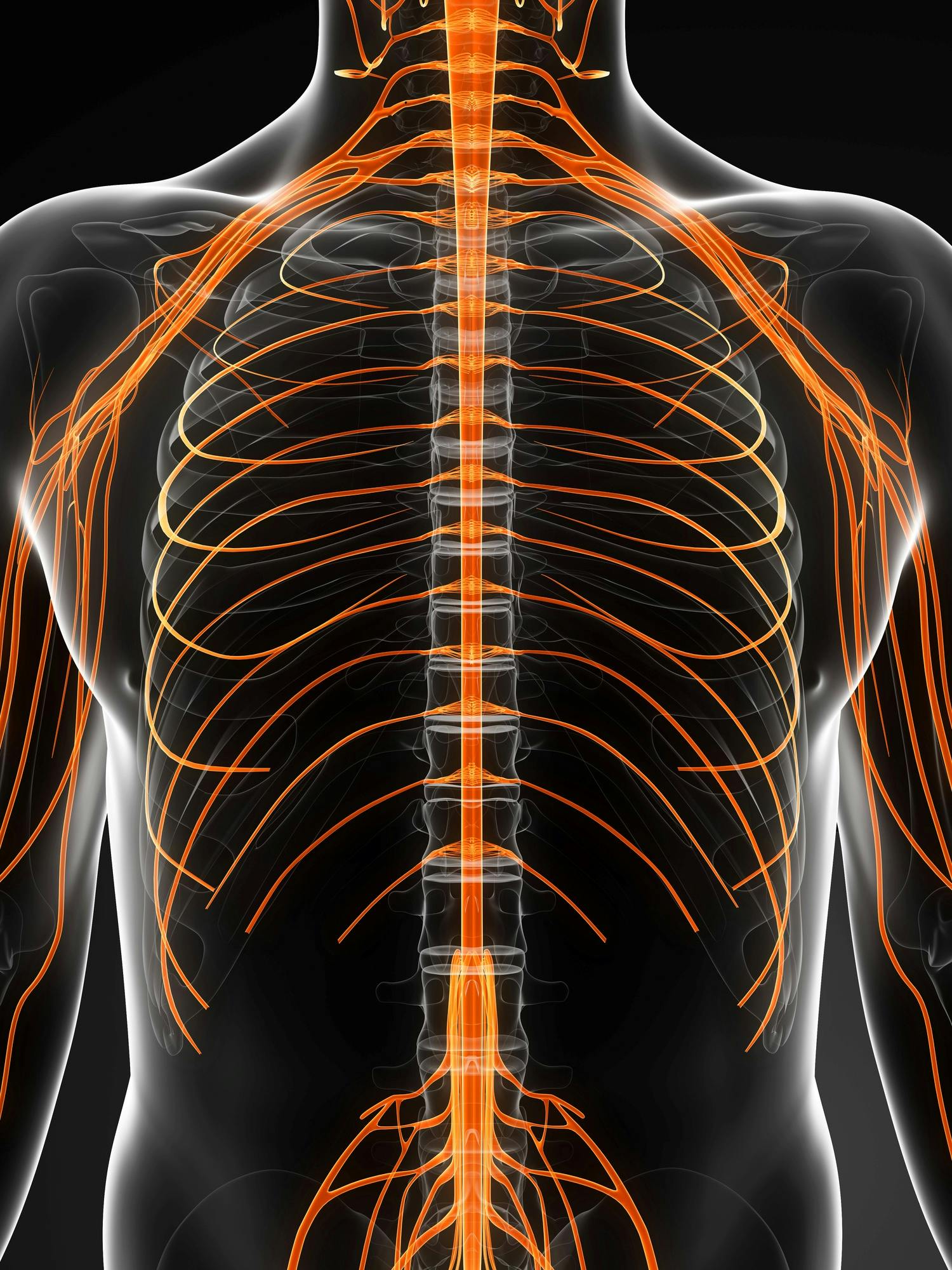
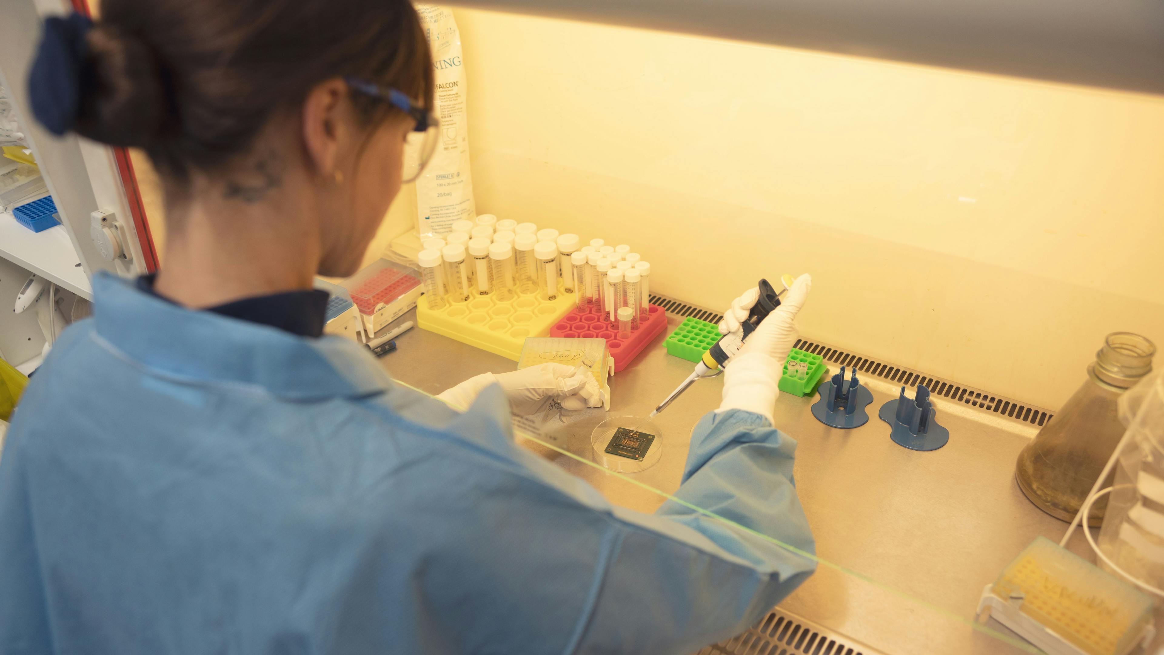
Microelectrode arrays, or MEAs, are widespread tools to increase our understanding of human physiology at the cellular level. They’ve long been used for in vitro cell interaction experiments that accelerate disease monitoring or drug screening.
However, most MEAs fall short in at least one of these aspects:
- They’re limited regarding the number of electrodes that can be accessed simultaneously. Or concerning the amount of information that can be extracted from a given sample.
- They cannot achieve the desired resolution (cellular precision or higher). This is due to the limited pitch of the electrodes.
- They don’t support high-throughput measurements. This slows down research.
- They’re passive devices without built-in circuitry. This requires complex external equipment.
- They cannot accommodate extra sensing & imaging modalities. These are needed to characterize complex cell behavior and interactions fully.
Thankfully, CMOS technology's miniaturization and integration potential makes it possible to make up for many of these inadequacies. CMOS MEAs pack a massive number of electrodes on one chip. That also includes signal processing, filtering and analog-to-digital conversion.
The result is a complete and compact system. It enables high-throughput cell interfacing with single-cell resolution and unprecedented signal quality.
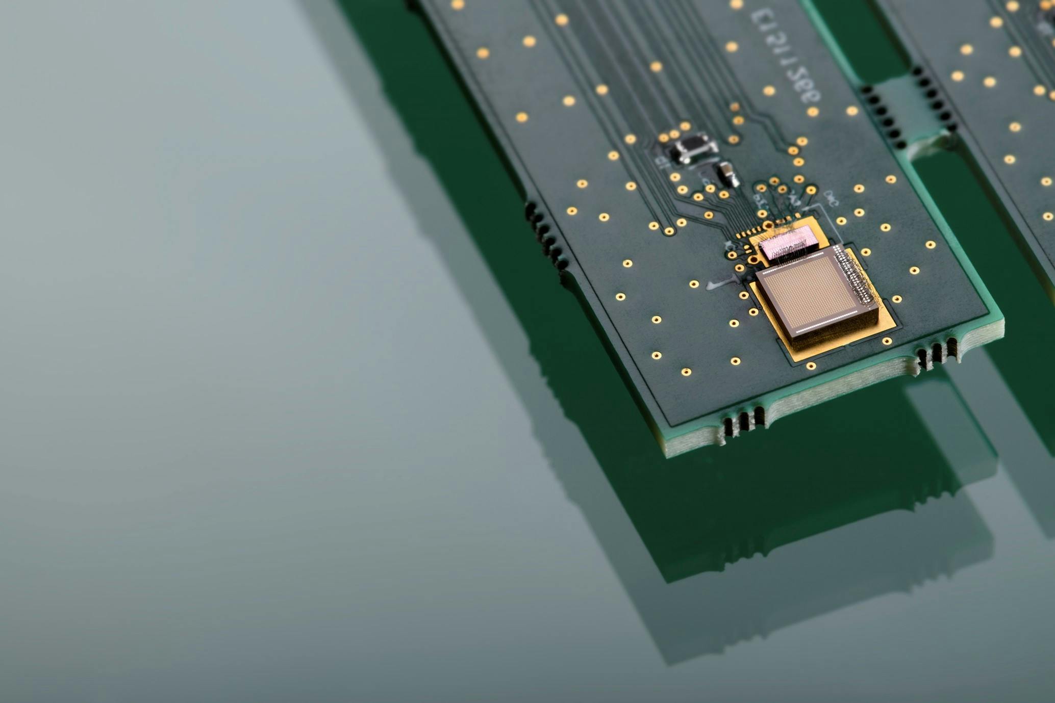
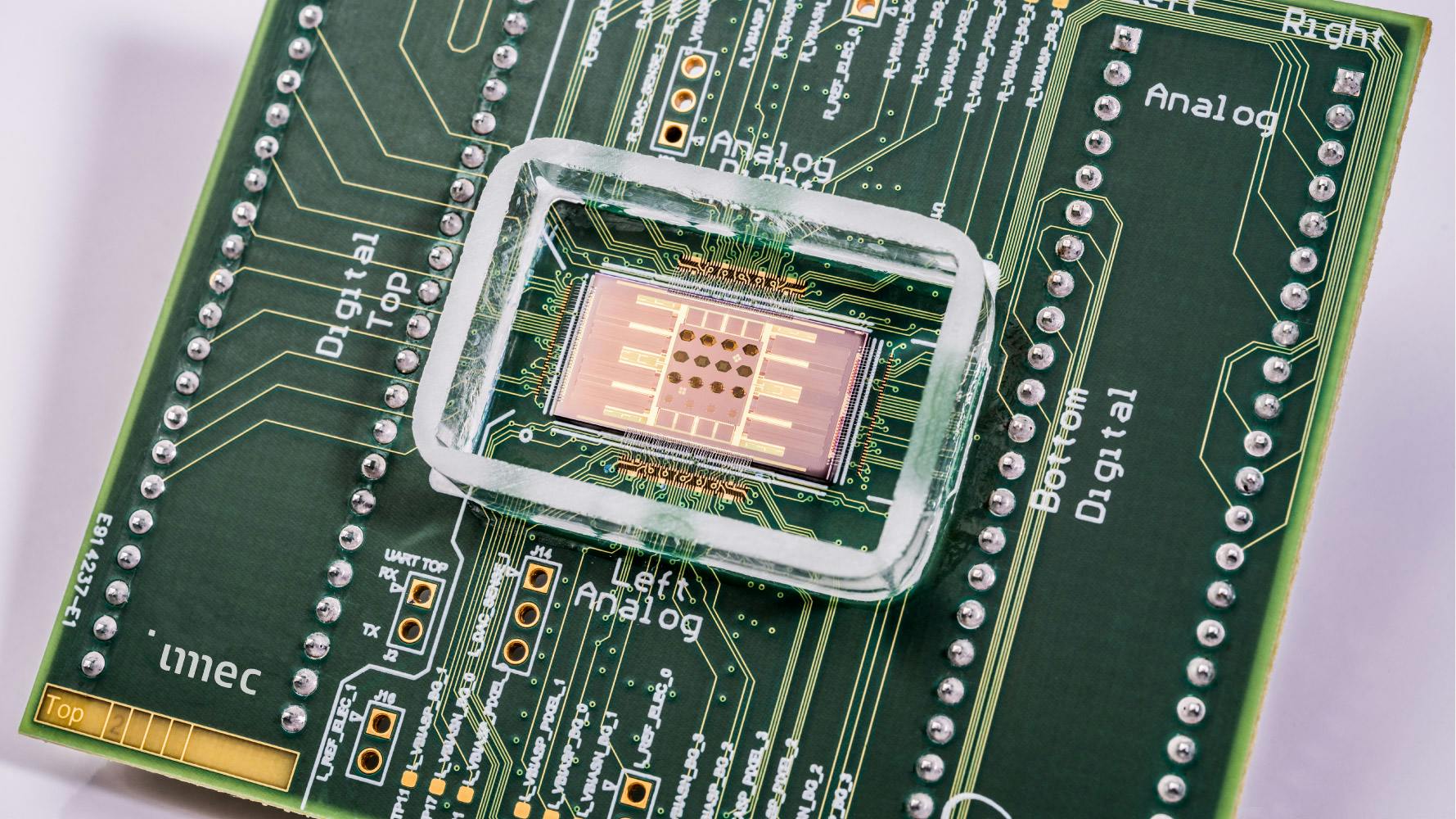
Imec’s CMOS MEA chip
The CMOS MEA chip developed by imec is an integrated solution for intra- and extracellular activity recording. It contains:
- 16,384 electrodes
- 1,024 low-noise read-out channels
Each electrode has a miniature pre-amplifier to improve the signal quality. The electrodes are grouped in 16 clusters that can be individually addressed. This makes it possible to run 16 experiments simultaneously. The MEA is covered with surface chemistry to make it compatible with cell cultures.
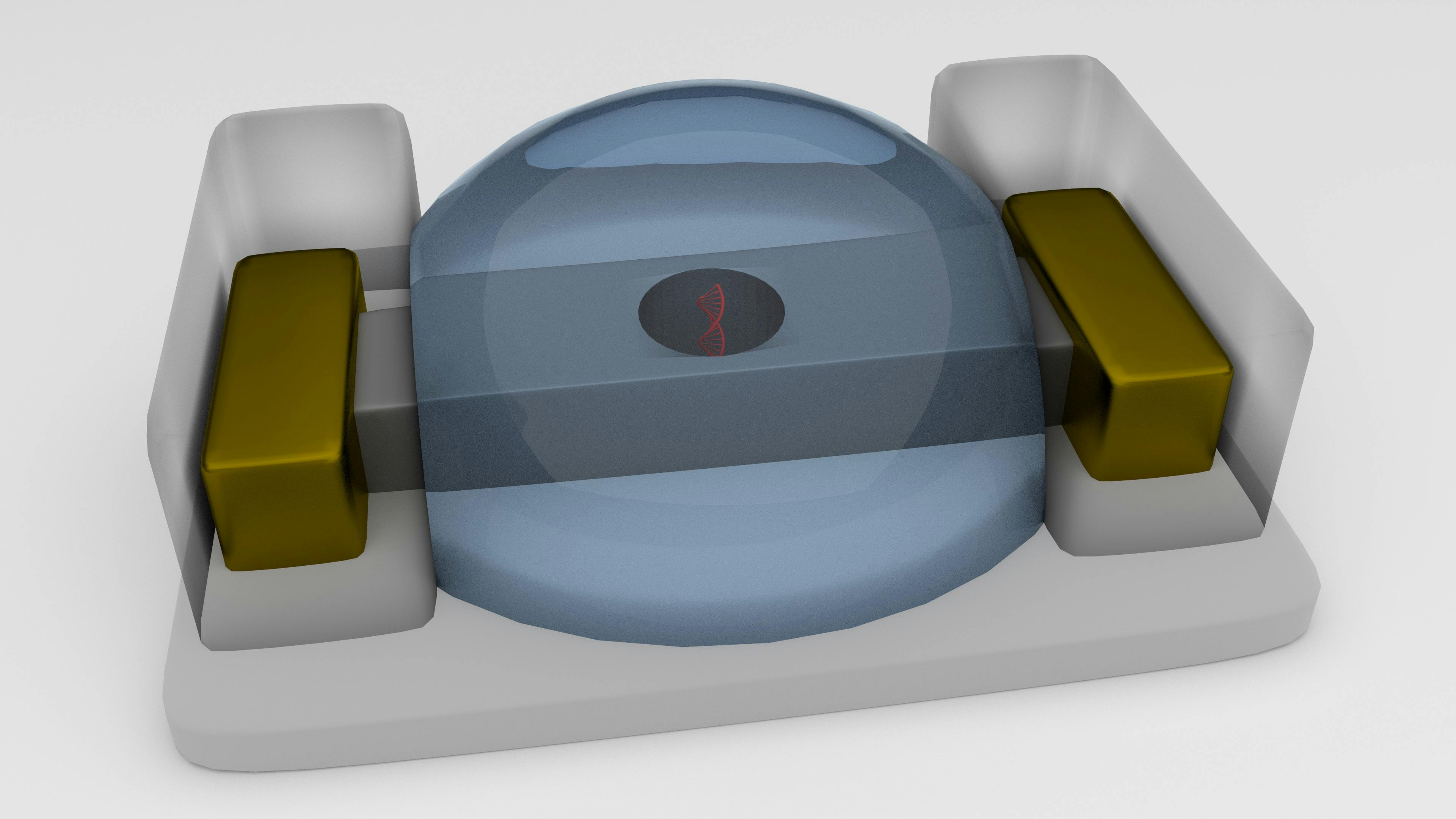
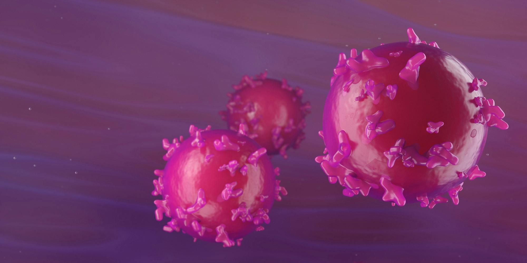
When a small voltage is applied to the cell via the electrode underneath, the cell membrane opens up. The molecules in the solution enter the cell. The voltage used and the size of these electrodes are so small that there’s no adverse effect on the cell. Experiments with human embryonic kidney cells 293 (HEK293) showed that a single cell can be transfected by the stimulation of one electrode (Duckert, 2021).
MEA-based single-cell electroporation allows an exact control of each cell's electroporation parameters. This enhances the precision of the technique. leads to more transfected cells in good condition. The results are increased yield and reproducibility and minimal toxic effects.
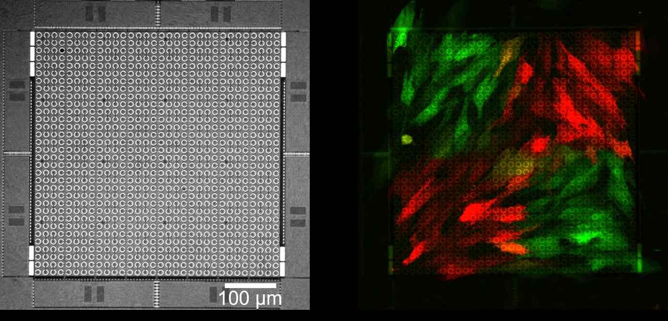
High-efficiency, spatially-resolved transfection of human fibroblasts on imec’s high-density microelectrode array. Left: close-up view of an electrode cluster with 1,024 circular, individually addressable electrodes. Right: fluorescence microscopy image of a cell transfected with an EFGP- (green) or mCherry- (red) encoding mRNA.
The reconfigurable on-chip circuits support six cell-interfacing modalities:
- extracellular/intracellular electrical activity recording
- voltage/current stimulation for cell excitation or local electroporation
- fast impedance monitoring to detect cell presence for optimal electrode selection
- single-cell impedance spectroscopy to give detailed information on electrode impedance, seal resistance and cell membrane impedance
The CMOS MEA chip can be packaged in a microfluidic well plate for cell culturing. Customized software enables stimulation, recording, programming of experiments and long-time follow-up of cells.
From CMOS MEA to organ-on-a-chip
Imec’s CMOS MEA can be used to precisely reprogram cells and differentiate them in a very controlled arrangement. In other words, they allow to build on-chip tissue models that very closely mimic the properties of actual organs. Their behavior can be registered over a long period.
The MEA is an essential part of imec’s organ-on-chip research activities that aim to improve biological models without resorting to animal testing. The current focus is on implementations such as:
- brain-on-a-chip technology to enhance our knowledge of the mechanisms of neurodegenerative diseases
- heart-on-a-chip technology
- modeling of the blood-brain barrier.
More about imec’s organ-on-chip research
Work with us
Want to examine the possibilities of imec’s CMOS MEA technology and tune it for your biomedical application?
Click the button below to get in touch