Photoacoustic spectroscopy and photoacoustic imaging
Photoacoustic-based medical imaging combines the resolution of light-based imaging and the depth penetration of sound-based imaging.
In 1880, Alexander Graham Bell discovered that certain materials emit sound waves when struck by pulses of light. Today's IC and photonics technology can turn this concept into unique chips for non-invasive medical imaging. Applications range from cortisol sensing to functional brain imaging. Complementing X-ray, MRI and ultrasound.
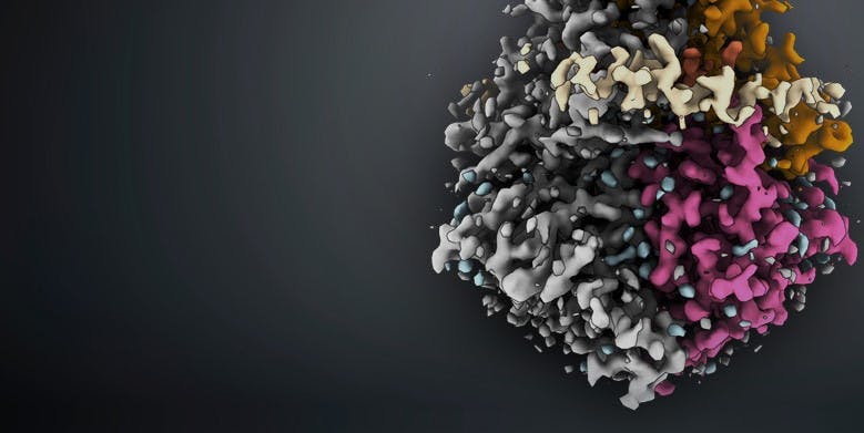
The technique uses no fluorescent labels or tags. Instead, it makes use of the different light absorption spectra of (distinct tissue types, such as) blood vessels, fat tissue etc. By illuminating the tissue at various wavelengths, different structures can be visualized simultaneously in one image.
Photoacoustic imaging (PAI)
This technique uses light sources with only one or a few wavelengths, depending on the absorption peak of the target structure or molecule. These are typically high-power pulsating light sources.
Photoacoustic spectroscopy (PAS)
Photoacoustics can be applied to identify biomolecules and their concentration based on their unique spectral signature. This application requires a tunable light source or one with a broad wavelength range.
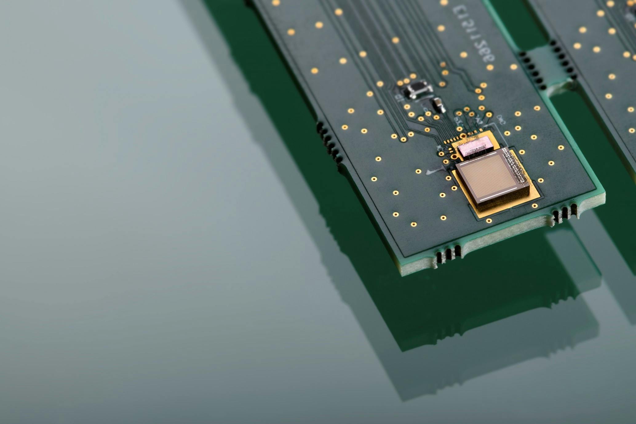
Light sources and microphones on chip
Imec’s silicon and silicon nitride integrated photonic platforms form the backbone of its photoacoustics systems-in-package. It comprises on-chip waveguides, ring resonators, spectrometers, couplers, etc. Based on these components, integrated light sources and microphones are realized towards high-performant and cost-effective photoacoustic solutions.
Imec developed integrated light sources for both PAI and PAS:
- dual-comb laser, based on a silicon nitride mode-locked laser
- silicon nitride waveguide with an indium phosphide laser on chip
- THz photoemitter array
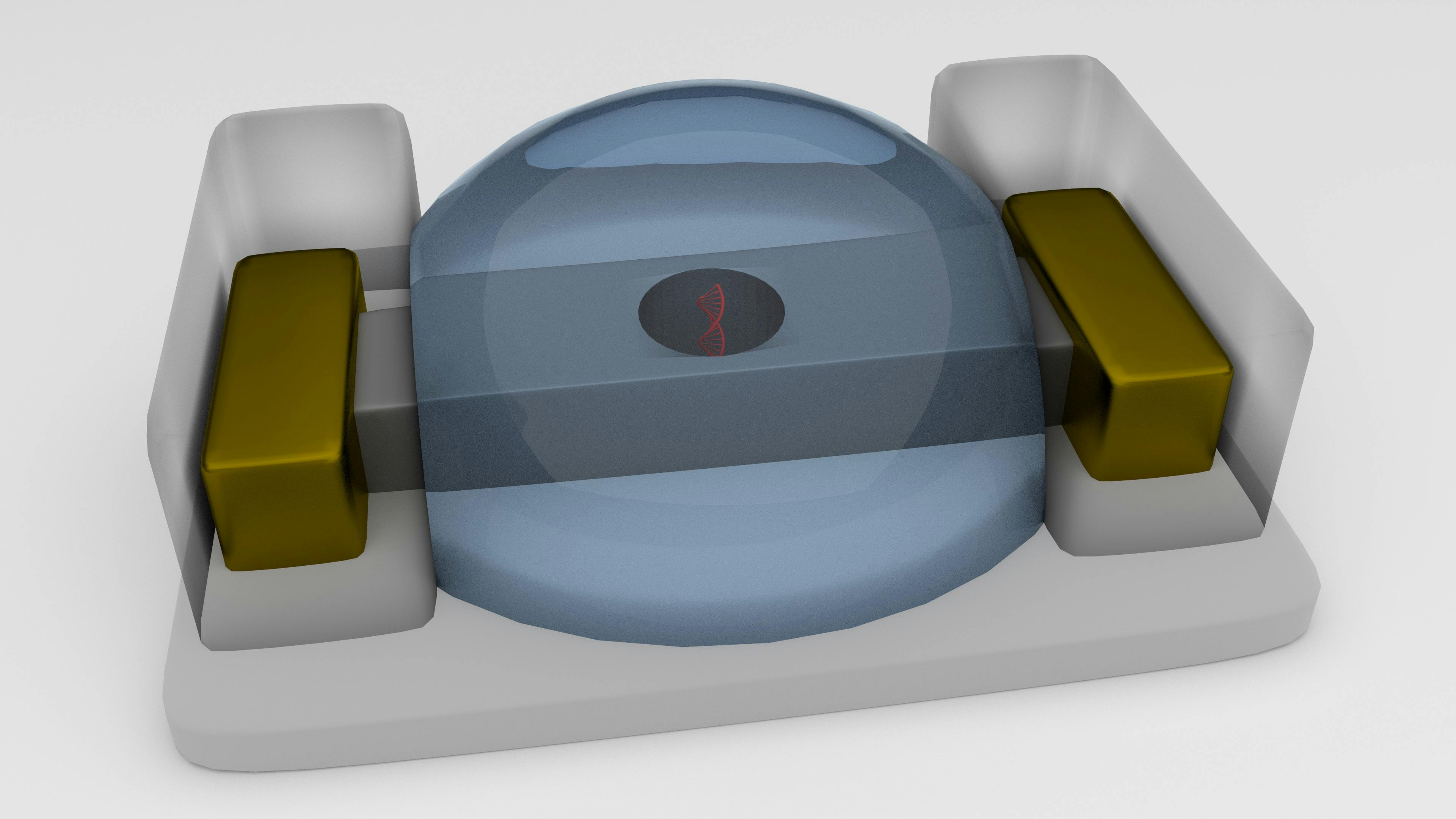
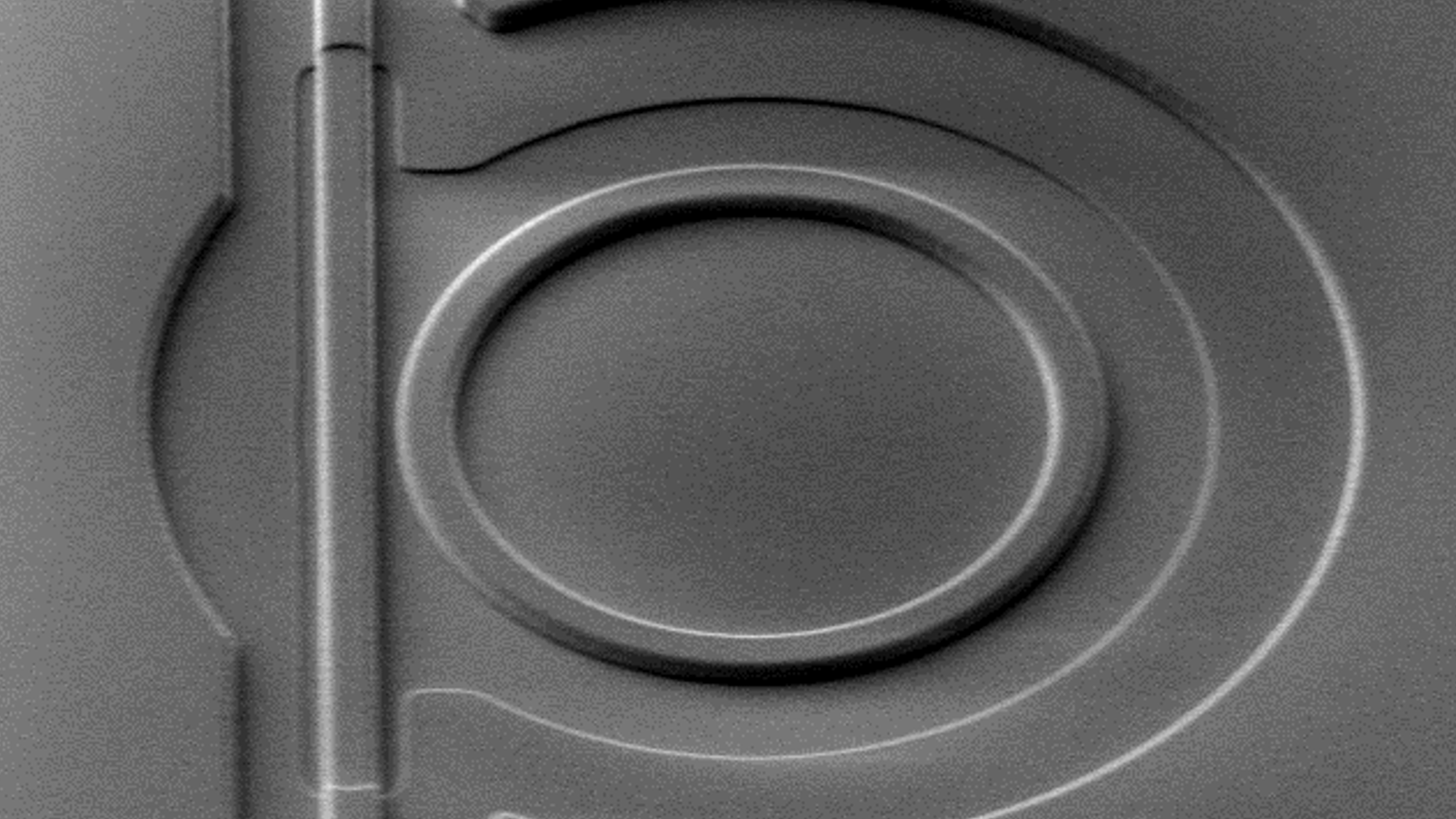
The generated sound waves can be detected by (an array) of microphones. The more sensitive the microphone, the deeper in the tissue one can listen. The optomechanical ultrasound sensor (OMUS) developed by imec consists of:
- sensitive silicon waveguide
- ring resonator
- free-standing membrane
The OMUS is recognized as a new benchmark within the photoacoustic imaging community. This is because of its exquisite performance in terms of sensitivity and noise – two orders of magnitude better than the state of the art.
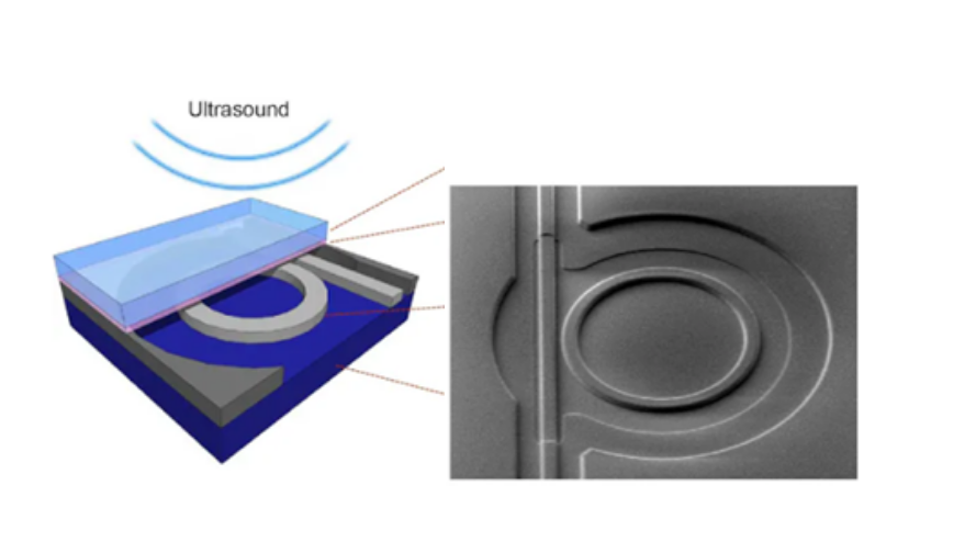
Imec’s optomechanical ultrasound sensor
Data analytics
The data generated by the integrated microphones are subsequently reconstructed to a high-resolution image, indicating the presence of specific molecules and structures. This requires advanced machine learning and data analysis.
Applications of photoacoustic spectroscopy and photoacoustic imaging
- functional brain imaging
- diabetes management
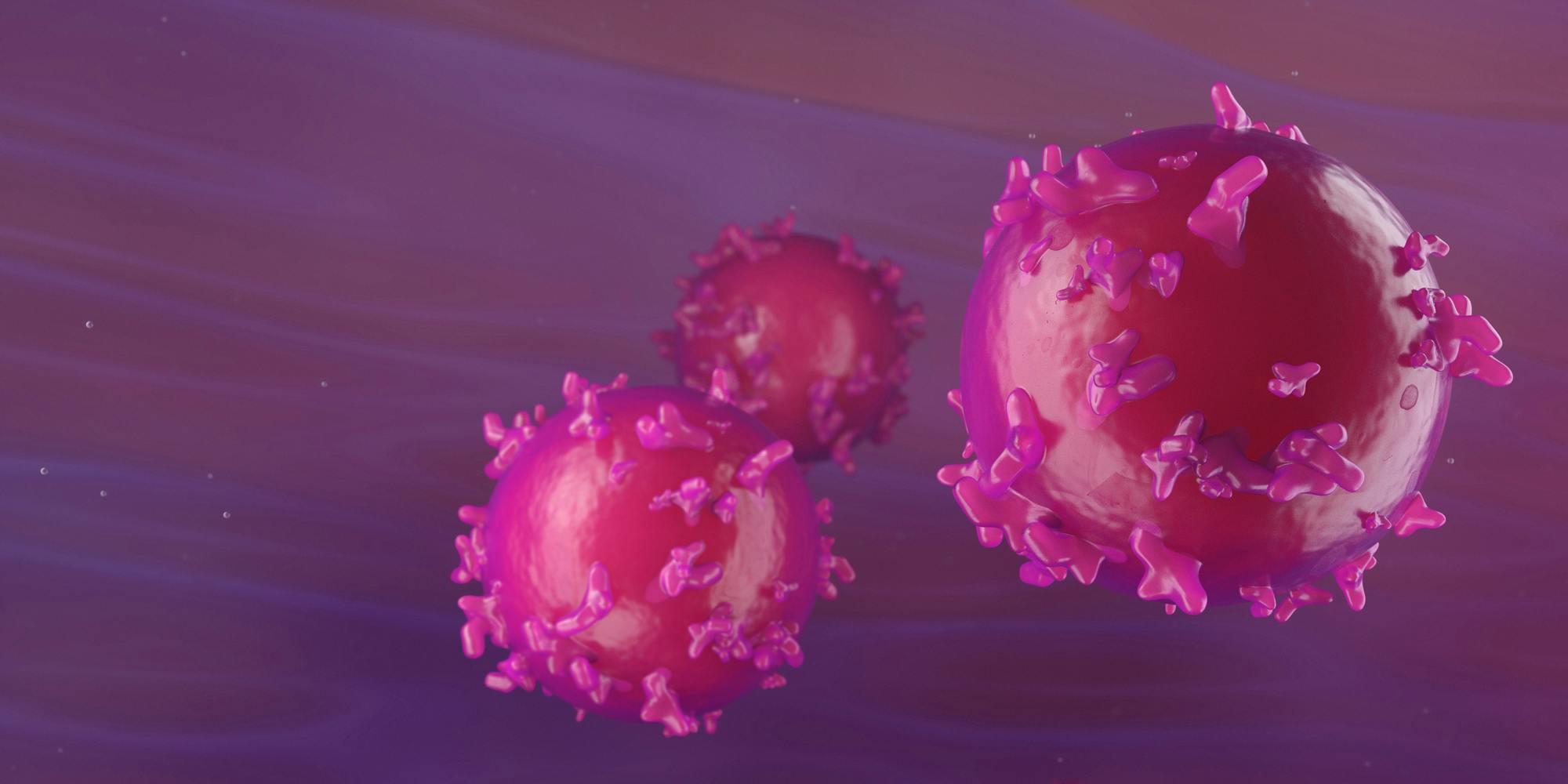
- melanoma detection
- breast cancer screening
- breath analysis to detect cancer
- inflammation monitoring such as in rheumatoid arthritis
- Blood vessel visualization after tissue grafting or to monitor wound healing
Click for an in-depth article on photoacoustics for biomedical applications
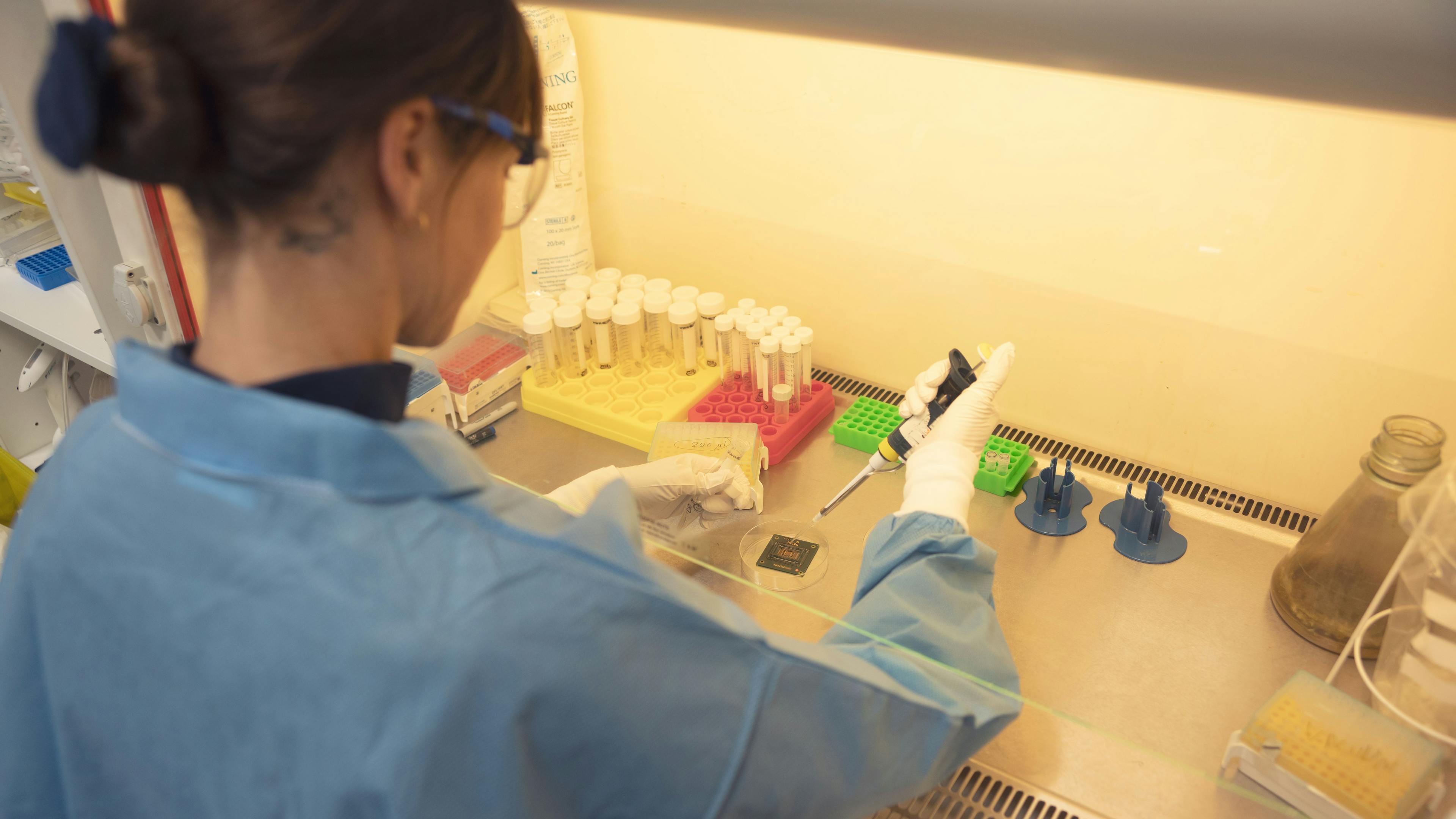
Imec is now building Proof-of-Concept demonstrators to enable collaborations with both clinical and industral partners towards clinical use of this exciting new technology. Are you working on a next-gen medical imaging system? Accelerate your innovation by leveraging our infrastructure, expertise and IP. We can develop a custom solution and support you all the way to prototyping and (chip) manufacturing.
Click the button below to get in touch.