AI-based Multimodal Image Analysis of Microvasculature on Chip
Gent | More than two weeks ago
Help create groundbreaking organ-on-chip technologies using AI-driven microvascular image analysis.
Using IMEC’s innovative microfluidic chip, 3D vessel-like networks can be grown. This perfusable vascular-on-chip platform has broad applications in the field of organ-on-chip, such as understanding diseases, drug testing, personalized medicine, and cell-based therapies (for more information see IMEC’s organ-on-chip technology [1]). However, there is a lack of non-invasive tools to characterize on-chip 3D vascular morphology and to assess other crucial parameters such as perfusion and oxygenation that greatly influence artery-capillary-vein development on the cell level and specification of endothelial cell lineage.
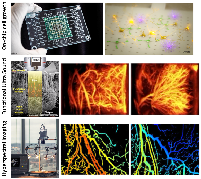
The objective of this PhD topic is to evaluate the growth and function of vascularization on chip using cutting-edge imaging technologies of IMEC such as Hyperspectral Microscopy, Lens-Free Microscopy, Functional Ultrasound Imaging, and Photo-Acoustic Imaging. Hyperspectral imaging (HSI) is an emerging biomedical imaging modality, which makes use of spectral signatures to identify specific substances, such as the measurement of tissue oxygenation and perfusion quantification in a non-invasive way. Lens-Free Holographic Microscopy (LFHM) is an emerging biomedical imaging modality, that removes the need for expensive and bulky optical lens components to acquire and visualize microscopy images while having comparable submicron optical resolution as traditional optical microscopes, larger field-of-view in one shot, and the capability to visualize samples in 3D volume. Functional Ultra Sound Imaging (fUSI) is a novel ultrasound-based technology that allows us to monitor local hemodynamics on a 10-micron scale yielding complex volumetric datasets (3D-in-time up to 100 μm3 voxel size) whose analysis is particularly challenging due to the high dimensionality. Photo-Acoustic Imager (PAI) combines light and sound to create an image based on the Photo-Acoustic (PA) effect – the property of certain materials to emit sound when struck by pulses of light and since hemoglobin has a very strong PA signature, it makes PAI ideal to image blood vessels and oxygen saturation.
Apart from imaging, accurate image processing methods are required to analyze and quantify vessel structures. Specifically, analysis of complex anatomical and 3D-in-time data requires: (1) accurate automatic segmentation and centerline extraction of the smallest blood vessels in each applied imaging modality, (2) AI-based 3D-in-time image registration between time instances and modalities in order to track the changes in vessels over time, and (3) near-to-real-time processing and visualization to allow for live laboratory experiments.
The Life Science department of IMEC focuses on interfacing hardware (sensors and actuators, microfluidics) and wetware (cells and tissues) using nanotechnology to create novel tools for personalized medicine. The Image Processing and Interpretation group of IMEC has been working on a number of biomedical and medical image processing and analysis applications and has long-standing experience in vascular image analysis. IPI also has close collaboration with IMEC-Solutions (developing HSI sensors/cameras and lens-free imagers for a wide variety of application fields such as agriculture, machine vision, medicine, remote sensing, biomedicine, etc.), Neuro-Electronics Research Flanders – NERF-IMEC-VIB-KU LEUVEN (developing the fUSI technology and conducting animal studies to detect brain activity through changes in blood vessel dynamics) and the IMEC Photonics Research Group (developing Photo-Acoustic imagers).
We are looking for a motivated Ph.D. candidate who is interested in pushing cutting-edge imaging technologies to the field of organ-on-chip production. You will: (1) conduct experiments to design the optimal imaging setup as a combination of various imaging modalities (HSI, LFI, fUSI, PAI), (2) develop novel AI-based image registration and analysis models to allow for fusion of data from various sensors, and (3) design methods for efficient parallel data processing to allow for near-to-real-time assessment of microvasculature.
[1] https://www.imec-int.com/en/articles/imec-presents-novel-organ-on-chip-platform-for-drug-screening
Required background: Computer Science or equivalent
Type of work: 70% modeling/simulation, 20% experimental, 10% literature
Supervisor: Hiep Luong
Co-supervisor: Yoke Chin Chai
Daily advisor: Danilo Babin
The reference code for this position is 2024-124. Mention this reference code on your application form.