Artificial Intelligence assisted Lens-Free Holographic Microscopy for improving mammalian fertility
Gent | More than two weeks ago
Design AI-based image processing methods for Lens-Free Holographic Microscopy to improve mammalian (e.g. human, bovine) fertility.
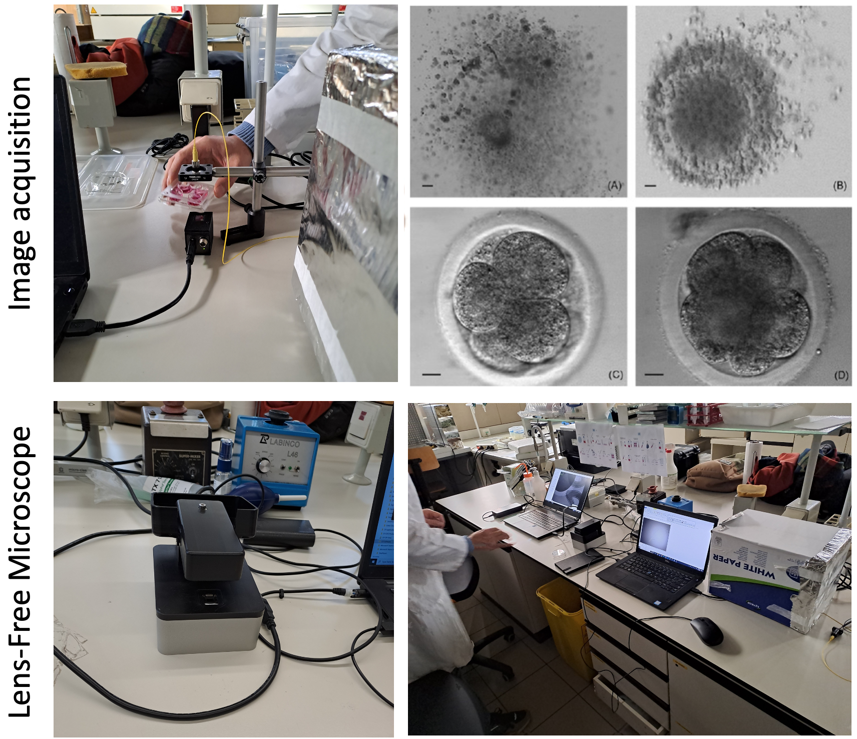
Lens-Free Holographic Microscopy (LFHM) is an emerging biomedical imaging modality, which removes the need for expensive and bulky optical lens components to acquire and visualize microscopy images. While having comparable submicron optical resolution as traditional optical microscopes, it is much smaller and less expensive, and captures images with an order of larger field-of-view in one shot, offering fast and high-throughput imaging capability. Recent developments in our research lab extended LFHM with the tomographic capability to visualize samples in 3D volume on a micrometer scale.1
The success of In-Vitro Fertilization (IVF) highly depends on the quality of oocytes and sperm used for fertilization. Ideally, only the highest quality cells are used for fertilization. Currently, the quality of oocytes is inspected visually using stereo microscopy and subjective rules to measure oocyte quality (brightness, granulation, contrast, size…), which yields moderate results. There is a clear need for objective algorithms to quantify oocyte quality and predict the outcome of fertilization. Stereo microscopy requires separate imaging of each oocyte complex (17 are placed together on the same Petri dish), prolonging imaging times and influencing the quality of cells in the process. Additionally, the produced image is a 2D projection of a 3D cell complex, resulting in a simplified view of the structure. Therefore, there is a clear need for using Lens-Free microscopy to image the oocyte complex on a wider field of view (the whole Petri dish can be imaged simultaneously) and to analyze the whole cell complex as a 3D structure to get better quality assessment and prediction.
The Integrated Imaging group of IMEC has vast experience in the development of imaging sensors and cameras for a wide variety of application fields (agriculture, machine vision, bio-medicine, remote sensing, etc.). The Image Processing and Interpretation group of IMEC has been working on a number of biomedical and medical image processing and analysis applications and has long-standing experience in the field. The Reproductive Biology Unit of the Faculty of Veterinary Medicine at Ghent University (the experimental partner) focuses on the interaction of embryos and gametes with their environment in different mammalian species and optimizes cryopreservation and culture techniques.
We are looking for a motivated Ph.D. candidate who is interested in establishing Lens-Free Imaging technology as the new state-of-the-art for In-Vitro Fertilization. You will: (1) conduct experiments to design the optimal 3D reconstruction method for in-vitro imaging (2) develop novel image reconstruction, processing, and analysis methods to quantify the quality of oocyte complex and sperm cells (3) design and implement the AI-based oocyte fertilization prediction method.
1 Luo et al. Vol. 28, No. 18, Optics Express 26935, 2020
Required background: Computer Science or equivalent
Type of work: 70% modeling, 20% experiments, 10% literature
Supervisor: Hiep Luong
Co-supervisor: Abdulkadir Yurt
Daily advisor: Danilo Babin
The reference code for this position is 2024-025. Mention this reference code on your application form.