Optical microscopy is an indispensable tool in biomedical research. Over the past decades, it has enabled researchers to visualize biological structures with remarkable precision, down to tens of nanometres, revealing processes at the level of individual cells and molecules.
But as we descend deeper into the biological tissue the sharp, high-resolution images obtained via optical instruments begin to lose their definition. In other words, today’s microscopes can be used for imaging close to the surface, but struggle to deliver high-resolution images at depth.
The reason lies in how light interacts with the complex, heterogeneous structure of living matter. As photons travel through tissue, they interact with different structures, such as cell membranes, organelle, and other microscopic features, which cause them to scatter. With each scattering event, photons deviate further from their original trajectory, gradually losing the directional information needed to form a sharp image. In result, the image is losing contrast and resolution.
For optical imaging, this presents a fundamental challenge as it limits how deep scientists can investigate living matter. One promising way to overcome this limitation is wavefront shaping, a technique that compensates for scattering by controlling how light enters and propagates through a medium. However, as living matter is constantly in motion, applying wavefront shaping in living tissue is far from straightforward. Researchers at imec and KU Leuven are therefore exploring how integrated photonics can make wavefront shaping faster and more scalable, paving the way for new imaging applications.
Wavefront shaping: turning fragmentation into focus
At first glance, the scattering of light inside biological tissue may appear hopelessly random. Yet beneath this apparent randomness lies an underlying logic: every photon's path is shaped by the structure of the material it travels through. If that structure can be understood, then, in principle, the distortion it causes can also be reversed.
Wavefront shaping builds on this principle. Instead of sending a beam of light into tissue, the incoming illumination wavefront is carefully tailored before it enters. In practice, this can be achieved using devices such as spatial light modulators (SLMs), for example via a liquid crystal device similar to displays (LCDs), which adjusts the properties of the incoming light pixel by pixel. This way, the complex scattering behaviour of the medium can be identified and compensated. What would normally dissolve into a diffuse glow is effectively pre-corrected, allowing the scattered light to refocus into a bright, well-defined spot.
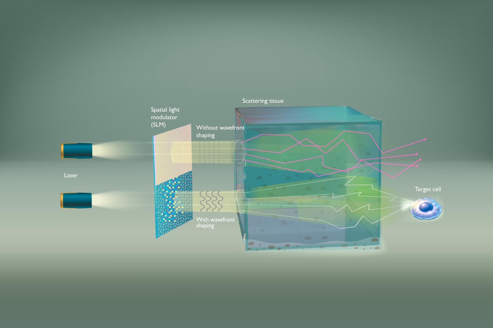
Illustration of the wavefront shaping principle. When no wavefront shaping is applied, the illumination beam starts to diffusely spread. By optimizing the incoming wavefront, wavefront shaping compensates for scattering and brings the light back into a focus.
The concept is powerful, but implementing wavefront shaping in real biological environments introduces a crucial challenge: speed. Living tissue is not static. Cells move, blood flows, and physiological processes continuously reshape the microscopic structure through which light propagates. As a result, the scattering pattern that wavefront shaping seeks to compensate for changes continuously over time. To remain effective, the correction applied to the incoming light must therefore be updated at comparable speeds, often on millisecond timescales or faster. Conventional wavefront shaping approaches, which rely on relatively slow modulation technologies, struggle to keep pace with these dynamics. This makes it difficult to maintain a stable focus deep inside living tissue.
Bringing wavefront shaping onto a chip
To address this speed limitation, researchers at imec and KU Leuven turned to integrated photonics, bringing wavefront control onto a photonic chip.
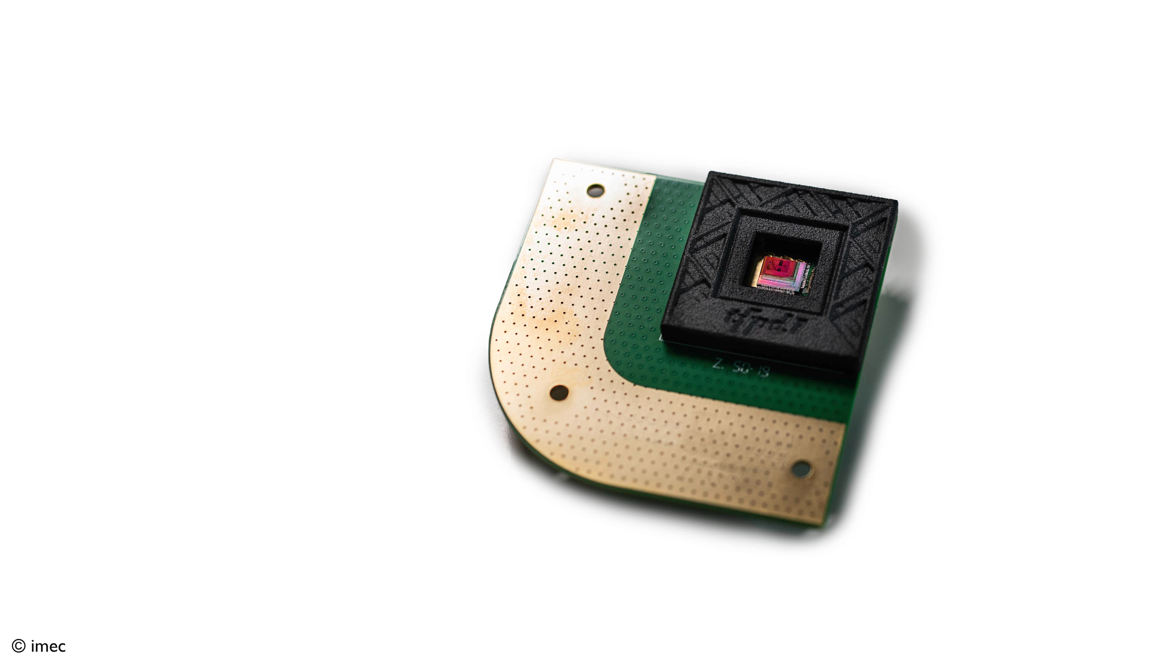
At the core of this concept are arrays of microscopic waveguides etched onto the chip, known as optical phased arrays (OPAs). These structures consist of many individually controlled light-emitting channels, whose phases can be tuned with high precision. By adjusting these phases in a coordinated way, the chip can shape the incoming illumination wavefront, effectively applying the corrections needed to compensate for scattering inside the tissue.
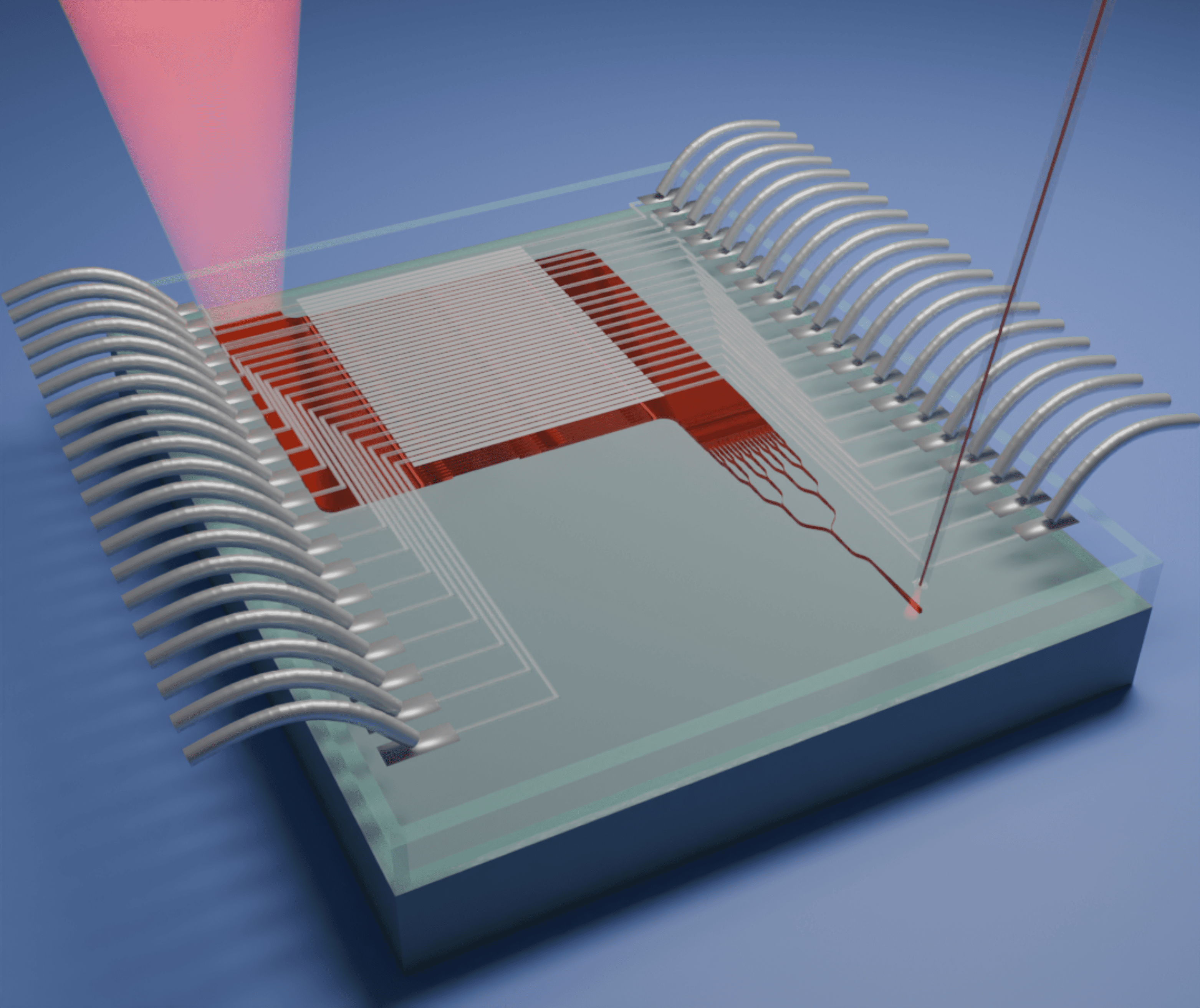
Illustration of a photonic chip employed as a wavefront shaper. Laser light coming from an external source enters the chip through an optical fiber, is modified as it travels through the chip, and is then emitted in a high-resolution wavefront modulation.
In a series of studies published in ACS Photonics [1,2], Nanophotonics [3], the researchers demonstrated that these integrated photonic chips can successfully perform wavefront shaping in strongly scattering media. The experiments showed that the chips could refocus scattered light into a well-defined spot with focus contrasts reaching about 90% of the theoretical maximum. This means that, even after light has been strongly scrambled, the chip can effectively compensate for scattering and bring light back into focus with almost maximum efficiency.
Beyond effective light control, the team also showed that the chip technology can operate at update rates of around 5,000 wavefront corrections per second, making it significantly faster than conventional liquid crystal-based modulators.
Together, these results demonstrate how integrated photonics can combine effective light control with the speed required for dynamic scattering environments. But its potential extends beyond performance alone. By integrating multiple optical functions on a single chip, wavefront shaping systems can become significantly more compact than conventional approaches based on liquid crystal modulators. At the same time, the chip-based platform makes it easier to combine wavefront shaping with additional functionalities, creating a flexible foundation for future optical imaging systems.
Towards practical imaging applications
These results highlight the potential of integrated photonics as a scalable platform for wavefront shaping. The next question is where its combination of speed, precision and compactness can have the greatest impact.
By actively controlling how light propagates through scattering media, wavefront shaping can restore focus in situations where image quality would otherwise rapidly deteriorate, opening the door to applications that are difficult or impossible to achieve with conventional optical approaches.
This is particularly relevant for fields such as neuroscience, where researchers rely on optical methods to study brain activity but are constrained by limited imaging depth. Extending that range, while preserving resolution, would allow them to observe biological processes that cannot currently be measured directly.
The same principle applies to biomedical diagnostics. Improving how light is guided through tissue could enhance the contrast and sensitivity of optical imaging techniques, supporting more accurate detection of diseases.
Another promising, and perhaps less intuitive, application area lies in multimode optical fibers. While optical fibers are best known for transmitting large amounts of data in telecommunications networks, their small diameter also makes them attractive as ultra-thin imaging probes that can reach locations deep inside the body that are impossible to access with conventional instruments. Yet this advantage comes with a challenge: as light travels through the fiber along many different paths, the optical signal becomes distorted, preventing the formation of a sharp image. By shaping the incoming light with an optical phased array, researchers can compensate for this distortion and reconstruct a sharp focus at the fiber's output. This could enable high-resolution imaging through a hair-thin fiber, significantly reducing the invasiveness of endoscopic procedures.
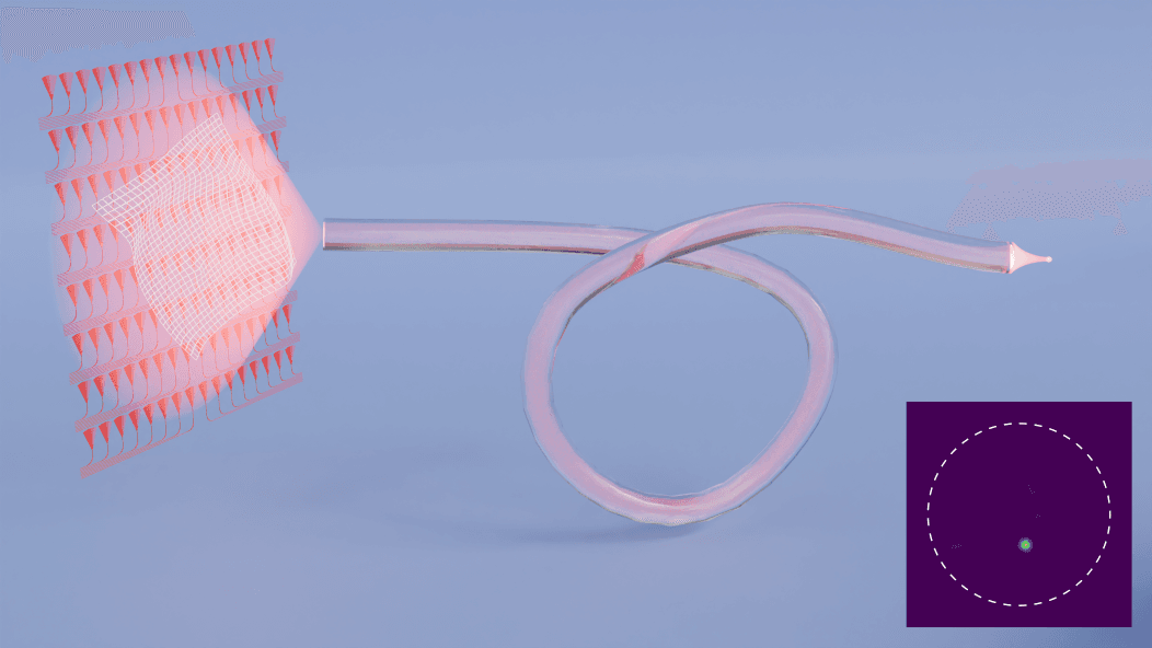
Illustration of wavefront shaping through a multimode fiber using integrated photonics: By shaping the incoming light, scattered light inside the fiber can be brought back into focus, enabling a high-resolution image.
The next steps: from proof of concept to broader application
Building on these emerging application opportunities, the development of integrated wavefront shaping continues to move forward. Further developments are underway to improve the underlying photonic platform, with a particular focus on increasing speed, scalability and robustness. At the same time, ongoing efforts are aimed at integrating more functionality onto a single chip, reducing system complexity and making wavefront shaping easier to deploy in practical settings.
What is clear though is that the successful combination of wavefront shaping and integrated photonics has been proven. Wavefront shaping already offered a way to recover information that scattering appears to erase. Integrated photonics now provides a route to doing so with greater speed, precision and compactness.
For optical imaging, that shift is significant. As one of the main barriers to deep tissue imaging, scattering has long constrained what can be observed with light. Integrated wavefront shaping could help overcome that limitation, making scientists able to see deeper and with greater precision into biological systems than ever before.
This article was originally published in Laser Focus World.
Want to know more?
[1] ‘Focusing through Scattering Media Using Integrated Photonics,’ F. Milojković et al., ACS Photonics, 2024
[2] ‘Integrated Photonics-Based Focusing through Multimode Fibers,’ F. Milojković et al., ACS Photonics, 2025.
[3] ‘Optical phased arrays for wavefront shaping in forward scattering media,’ F. Milojković et al., Nanophotonics, 2025.